Shoulder Muscles Diagram Posterior : Ever-Green Massage Therapy: May 2011. The shoulder muscles are associated with movements of the upper limb. Anterior part of the deltoid: The rotator cuff is a made up of four muscles in the shoulder, connecting the humerus to the scapula. 7.16 posterior muscles of the shoulder and arm. These muscles form the outer shape of the shoulder and underarm.
Posterior view of human muscular system. Each deltoid muscle has three heads, or distinct parts: They are shown in the image below. Posterior band of the ighl. Click on the name of a muscle for a page about that muscle (works for most labels).
Anatomy of the Shoulder - Part 3 (Muscular Structures) - MUJO from www.mujofitness.com Anterior graphic of the shoulder. Summary of the structure of the posterior shoulder muscles. The shoulder muscles include skeletal muscles that are attached to the head of the humerus which performs various direct and indirect functions of the shoulder joints. Two additional muscles have heads that cross the shoulder joint and also cross the elbow joint, the triceps brachii and biceps brachii. Important muscular spaces of shoulder. Left posterior basal segmental bronchus. Learn their origins/insertions, functions & exercises. Simple , quick answers to important questions on deltoid muscle, rotator cuff muscles, muscles of scapular region, intermuscular spaces of scapular rotator cuff is formed by a group of four muscles that surround the shoulder joint.
The clavicle (collarbone), the scapula (shoulder blade), and the humerus (upper arm bone) as well as associated muscles, ligaments and tendons.
Nine muscles cross the shoulder joint. Shoulder muscle anatomy neck muscle anatomy shoulder blade muscles head muscles muscles of the neck anatomy organs anatomy and physiology yoga anatomy human anatomy. Unidirectional posterior shoulder instability is much less common than anterior instability, however it should be strongly suspected in those high risk group of athletes with posteroir shoulder pain and/or clicking. Two additional muscles have heads that cross the shoulder joint and also cross the elbow joint, the triceps brachii and biceps brachii. Learn their origins/insertions, functions & exercises. Crosses brachial artery lateral to medial. With cross body stretching, people will often why is this relevant? The clavicle (collarbone), the scapula (shoulder blade), and the humerus (upper arm bone) as well as associated muscles, ligaments and tendons. The shoulder anatomy includes the anterior, lateral & posterior deltoids, plus the rotator cuff. General anatomy and the musculoskeletal system: Shoulder muscles and shoulder tendons. Important muscular spaces of shoulder. The approach also was used in 2 cases with fractures involving the scapular.
The anterior, lateral and posterior deltoid heads. The approach also was used in 2 cases with fractures involving the scapular. Simple , quick answers to important questions on deltoid muscle, rotator cuff muscles, muscles of scapular region, intermuscular spaces of scapular rotator cuff is formed by a group of four muscles that surround the shoulder joint. Want to learn more about it? Posterior muscles in the body.
File:Shoulder joint.svg - Wikipedia from upload.wikimedia.org The shoulder muscles are associated with movements of the upper limb. The anterior, lateral and posterior deltoid heads. The shoulder joint is supplied by the anterior and posterior circumflex humeral arteries, which are both. The human shoulder is made up of three bones: Muscles of the shoulder can be subdivided into a variety of groups depending on origin, topography, function or innervation. As a group, they are responsible for stabilizing the shoulder joint. 7.24 linear diagram of brachial plexus and branches. The treatment involves a combination of skilled therapy and surgery for optimal outcome.
Here we explain the major muscles of the human body.
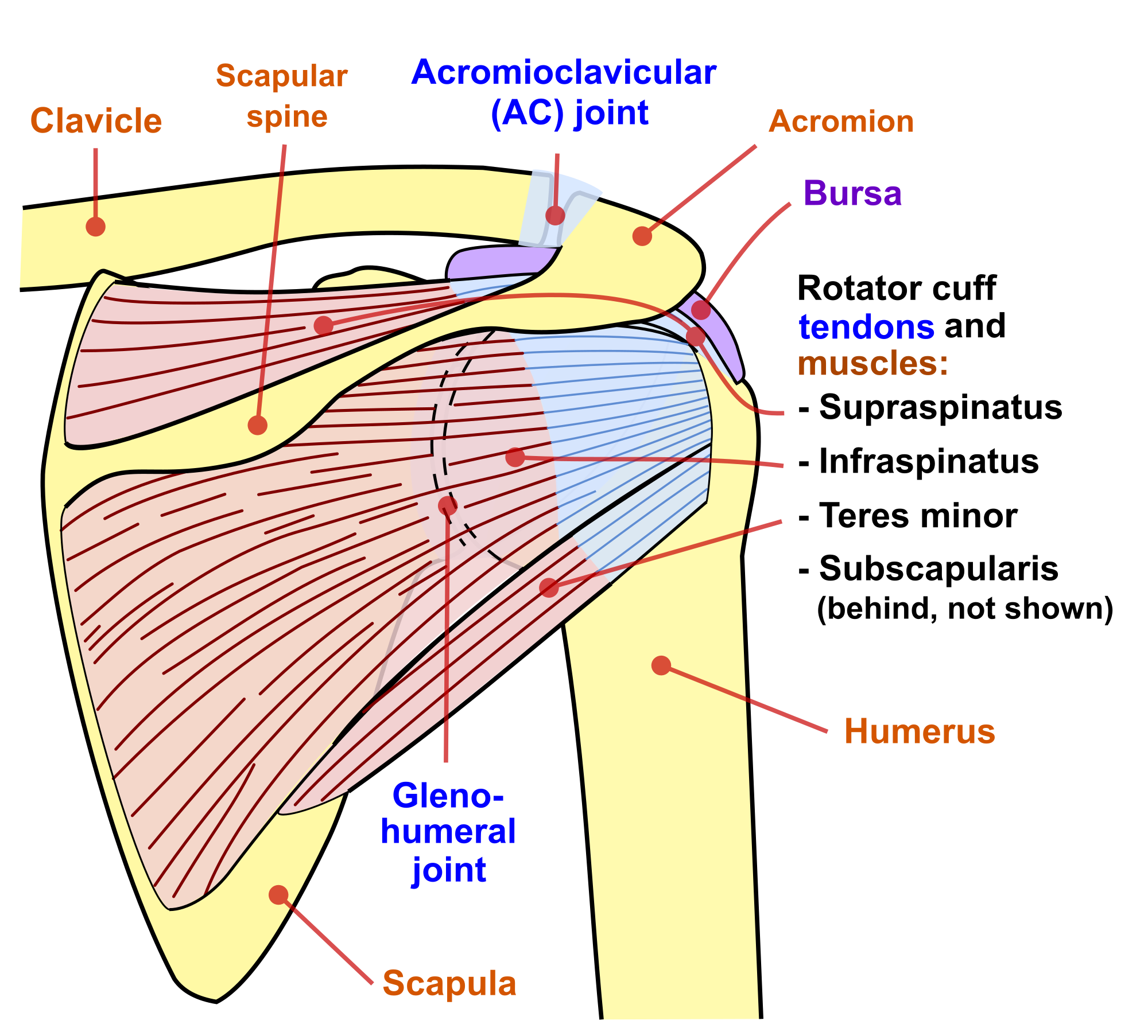
The first set, the shoulder's external rotators, includes the teres minor and the infraspinatus as well as the posterior part of the deltoid. Anterior part of the deltoid: The posterior muscles of the shoulder: Infraspinatus and teres minor tendon. This muscle diagram is interactive: Extends and laterally rotates the arm. Important muscular spaces of shoulder. Typically accompanies shoulder girdle downward rotation. Posterior band of the ighl. Superficial muscles of the posterior forearm: The shoulder has about eight muscles that attach to the scapula, humerus, and clavicle. Learn vocabulary, terms and more with flashcards, games and other study tools. Flexes and medially rotates arm;
The approach also was used in 2 cases with fractures involving the scapular. The human shoulder is made up of three bones: The shoulder anatomy includes the anterior, lateral & posterior deltoids, plus the rotator cuff. The posterior muscles of the shoulder: Typically accompanies shoulder girdle downward rotation.
Deltoideus | Anatomy Study Buddy from anatomystudybuddy.files.wordpress.com The posterior scalene muscles, located on the lower sides of the neck, ipsilaterally bend the neck to the side and elevate the second the back contains the origins of many of the muscles that are involved in the movement of the neck and shoulders. Learn their origins/insertions, functions & exercises. There are anterior muscles diagrams and posterior muscles. Related online courses on physioplus. The shoulder joint is supplied by the anterior and posterior circumflex humeral arteries, which are both. The shoulder muscles are associated with movements of the upper limb. The shoulder has about eight muscles that attach to the scapula, humerus, and clavicle. Superficial muscles of the posterior forearm:
The human shoulder is made up of three bones:
Related online courses on physioplus. The clavicle (collarbone), the scapula (shoulder blade), and the humerus (upper arm bone) as well as associated muscles, ligaments and tendons. Anterior part of the deltoid: The rotator cuff is a made up of four muscles in the shoulder, connecting the humerus to the scapula. Muscles allow us to move by pulling on bones. Muscles of the shoulder can be subdivided into a variety of groups depending on origin, topography, function or innervation. Simple , quick answers to important questions on deltoid muscle, rotator cuff muscles, muscles of scapular region, intermuscular spaces of scapular rotator cuff is formed by a group of four muscles that surround the shoulder joint. Posterior part of the deltoid: This muscle diagram is interactive: Shoulder muscles and shoulder tendons. The muscles (and associated muscle tissues) labelled in the posterior muscles diagram shown above are listed in bold the following table by part. As a group, they are responsible for stabilizing the shoulder joint. Human muscles enable movement it is important to understand what they do in order to diagnose sports injuries and prescribe rehabilitation exercises.
Related online courses on physioplus shoulder muscles diagram. Posterior view of human muscular system.
Share :
Post a Comment
for "Shoulder Muscles Diagram Posterior : Ever-Green Massage Therapy: May 2011"


Post a Comment for "Shoulder Muscles Diagram Posterior : Ever-Green Massage Therapy: May 2011"